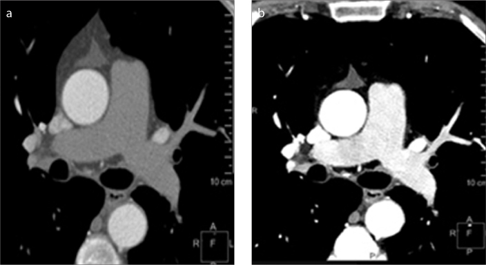
Figure 3. a, b.
Salvage of a suboptimal pulmonary embolism study. Conventional polyenergetic CT image (a) obtained for evaluation of pulmonary embolism with poor vascular enhancement due to contrast extravasation. Virtual 40 keV monoenergetic image (b) shows significantly improved enhancement permitting evaluation of the pulmonary arteries, obviating the need for a repeat contrast injection.

