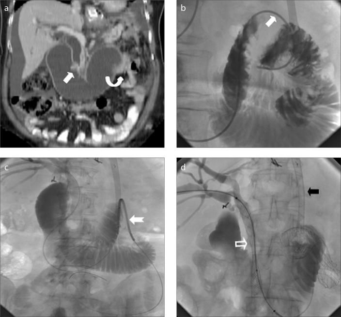
Figure 2. a–d.
A 53-year-old female with gastrectomy and Roux-en-Y anastomosis. Coronal reformatted abdominopelvic CT image (a) demonstrates biliary and afferent loop dilatation due to obstructive recurrent tumors at ampulla (arrow) and distal end of afferent loop (curved arrow). Fluoroscopic images (b–d) obtained during transhepatic metallic stenting show traversing obstructed afferent loop segment (arrow) with a catheter, placement of metallic stent (notched arrow) over an exchange wire and metallic biliary stenting (hollow arrow), respectively. Note previously placed esophagojejunal metallic stent (black arrow).

