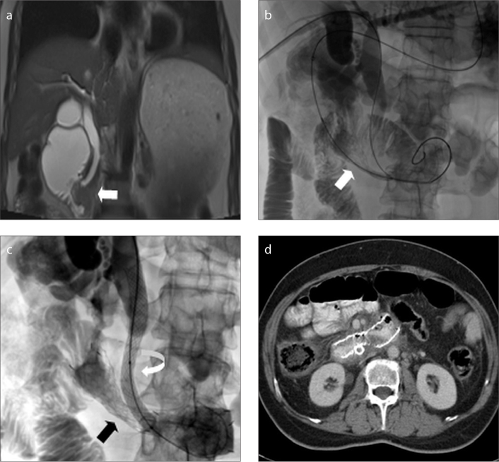
Figure 3. a–d.
A 55-year-old patient with pancreatic cancer. Coronal magnetic resonance imaging (a) shows an infiltrative mass causing both biliary and duodenal obstruction (arrow). Fluoroscopic images (b–c) obtained during simultaneous biliary and duodenal stenting demonstrate transoral placement of duodenal stent (arrow) and transhepatic placement of metallic biliary stent (curved arrow), respectively. Expanded duodenal stent is also shown (black arrow). Axial CT image (d) obtained 56 weeks after stenting shows patent stent at duodenum.

