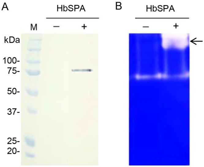
Fig 3. Transient expression of HbSPA in N. benthamiana.

Intercellular fluids were isolated from N. benthamiana leaves infiltrated with A. tumefaciens GV3101 carrying the pJL3-P19 as a control (- HbSPA), a mixture of A. tumefaciens strains carrying pGD-HbSPA and pJL3-P19 (+ HbSPA). (A) Samples were analyzed by SDS-PAGE followed by Western blot with HRP conjugated anti-His monoclonal antibody. Lane M represents the protein standard and the numbers on the left indicate the size of molecular weight markers. (B) Samples were analyzed by zymogram in-gel protease assay. Samples were loaded on a SDS-PAGE containing 0.1% of gelatin. After electrophoresis, proteins were renaturated in the gel and the protease activity was detected after a 2-h incubation required for gelatin degradation. The clear and distinct bands against the blue background on the gel corresponding to the protease activity associated with the presence of the HbSPA protein was indicated by an arrow.
