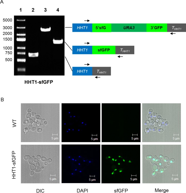
Fig 3. PCR analysis of tag-HHT1 yeast strains and their subcellular localizations.
(A) HHT1-sfGFP strain. The genomic DNA PCR data shows in the left panel. Lane 1: DNA ladder marker. Lane 2: the original strain yRH182. Lane 3 and 4: the pop-in and pop-out strain. (B) The subcellular localizations of HHT1-sfGFP. The yRH182 (upper) is the wild-type yeast strains without modification; the tagged strains HHT1-sfGFP are shown at the bottom. The images were obtained under Plan-Apochromat 63×/1.40 oil (Zeiss 5 Live).

