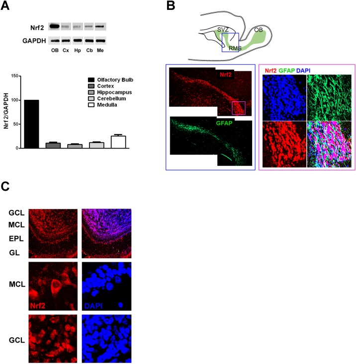
Fig 1. Nrf2 is highly expressed in olfactory bulb interneurons.
(A) Representative immunoblots and quantification of Nrf2 protein levels in adult murine brain (n = 4). Values were normalized by GAPDH and expressed as mean percentage (and S.E.M.) compared to olfactory bulb (OB). Cx, cortex; Hp, hippocampus; Cb, cerebellum; Me, medulla. (B) Schematic showing the rostral migratory stream (RMS), the route followed by neuroblasts originating in the sub ventricular zone (SVZ) to reach the olfactory bulb (OB). Immunohistochemistry showing the expression of Nrf2 in the RMS at low magnification (left) and high magnification (right). The boxed area indicates the regions shown in the immunostaining. (C) Immunohistochemistry showing the distribution of Nrf2 in the various regions of the olfactory bulb (upper panel). GCL: granule cell layer; MCL: mitral cell layer; EPL: external plexiform layer; GL: glomerular layer. The higher magnification panels show Nrf2 subcellular localization in mitral cells (MCL) and granule cells (GCL).

