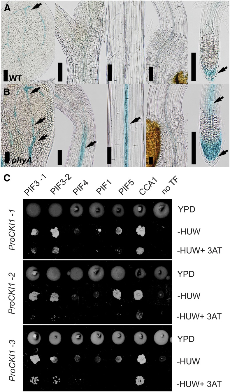
Figure 2.
CKI1 acts downstream of phyA-mediated signaling. A and B, In contrast to wild type, far-red light is unable to inhibit CKI1 expression in phyA (see also Fig. 1). To avoid possible bias due to developmental stage-specific changes, etiolated wild type and phyA carrying ProCKI1:uidA construct were treated with continuous far-red light (50 μmol m−2 s−1) immediately after the dark adaptation phase (4 d in darkness, 2 d on far-red). While in phyA, the CKI1 signal is still clearly detectable (arrows), only residual CKI1 activity can be detected in wild type, mostly in the vascular tissue of the cotyledons and in the LRC (arrows). Scale bars: 100 μm. C, Interactions between light-associated TFs and selected fragments of the CKI1 promoter (ProCKI1-1, ProCKI1-2, and ProCKI1-3) enriched in phyA-regulated motifs (see Supplemental Table S1 and Supplemental Fig. S5), identified by Y1H. Growth of yeast clones carrying the HIS3 reporter under the control of ProCKI1-1, ProCKI1-2, or ProCKI1-3 and selected TFs from the REGIA-REGULATORS collection was recorded after incubation for 6 d on either vector-selective media, interaction-selective media lacking His, uracil, and Trp or interaction-selective media supplemented with 10 mm 3AT, a competitive inhibitor of the HIS3 gene product. Only those yeast clones growing under the latter (more stringent) interaction-selective conditions were considered to be carrying interactors. PIF3 (both of the two independent clones available in the collection) specifically recognizes ProCKI1-1 and ProCKI1-2, while CCA1 interacts with all the fragments tested. HUW, His, uracil, and Trp; YPD, vector-selective media.

