Figure 3.
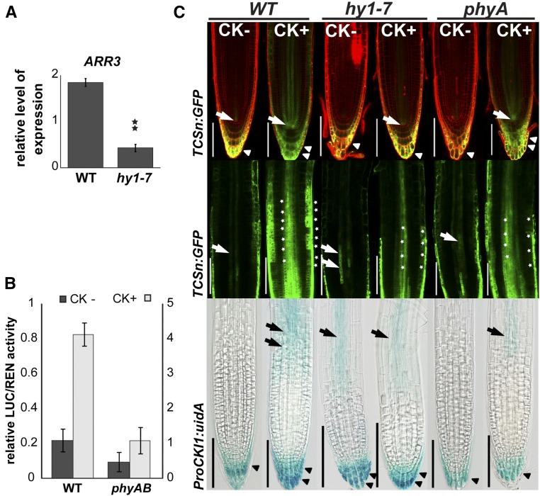
hy1-7 exhibits disturbed MSP activity and attenuated cytokinin sensitivity. A, Relative quantification by qRT-PCR of ARR3 expression in 6-d-old wild-type and hy1-7 seedlings grown under short-d conditions. Data were normalized to UBIQUITIN 10. Statistically significant difference at α = 0.01 is shown (**; n = 3). B, Relative expression of the cytokinin-responsive MSP reporter (TCS:LUC) in protoplasts from suspension cultures of wild-type and the phyA phyB mutant line; error bars show sd. Impaired MSP signaling in phyA phyB is apparent in both control (values on y axis on the left) and cytokinin-induced (0.1 μm BA) protoplasts (values on y axis on the right). C, Disturbed MSP signaling and inducibility of CKI1 expression in root tips of 6-d-old seedlings by cytokinin (0.1 μm BA). Upper row: Expression of cytokinin-responsive MSP reporter TCSn:GFP in the root apical meristem. In the absence of exogenous cytokinins, MSP activity in wild type is detected in the stele close to the quiescent center (arrow) and in the LRC and columella (arrowhead). In hy1-7 and phyA, the MSP activity in the stele close to the QC center is missing. In hy1-7 and partially also in phyA, the TCSn:GFP signal is decreased in LRC and almost missing in the cells of columella. In wild type, exogenous cytokinin treatment strongly up-regulates the TCSn:GFP signal in the stele, LRC (double arrowhead) and columella. In contrast, only modest cytokinin-mediated up-regulation is apparent in LRC (double arrowhead) and columella of phyA, and only very weak increase of MSP activity is detectable in case of cytokinin-treated hy1-7 (single arrow/arrowhead). PI contrastaining was used (red signal). Middle row: MSP activity as measured by TCSn:GFP expression in the root including proximal meristem and transition zone of wild type, hy1-7, and phyA. In comparison to wild type (single arrow), both mutants show increased activity of MSP signaling in the root vasculature including the transition zone (double arrow) in the absence of exogenous cytokinins. However, both phyA and particularly hy1-7 show only weak increase in MSP activity upon exogenous cytokinin treatment in the vascular tissue and epidermis (*). Bottom row: Disturbed cytokinin inducibility of CKI1 as shown by ProCKI1:uidA activity. Cytokinin up-regulates expression of CKI1 in the root transition zone (arrow) and columella/LRC (double arrowhead) of wild type, but not in the cytokinin-insensitive hy1-7 line; cytokinin only partially activated CKI1 in the transition zone and columella/LRC of phyA. Note the higher level of expression in the vasculature of the transition zone and in the columella/LRC in hy1-7 in the absence of exogenous cytokinin. In phyA, the CKI1 expression is slightly decreased in the LRC (arrowhead) in the absence of cytokinins. Scale bars: 100 μm.

