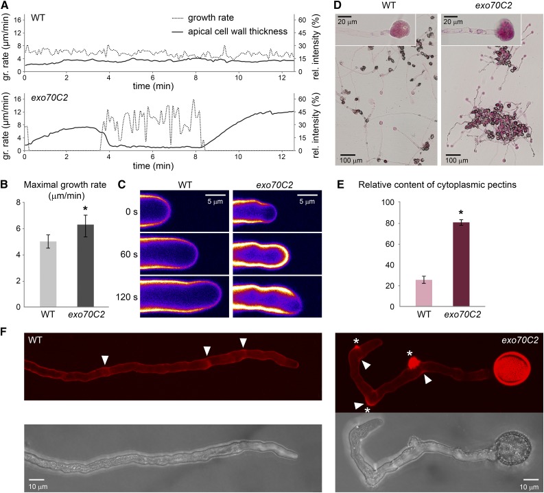
Figure 4.
Growth rate and cell wall characteristics of exo70C2 and wild-type (WT) pollen tubes. A, Growth rate of typical wild-type and exo70C2 pollen tubes correlated with cell wall thickness (Calcofluor White fluorescence) at the tube apex. Images for measurement were captured at intervals of 5 s. B, The averaged maximal growth rate of exo70C2 is significantly higher than that of the wild type (sd is displayed: *, P < 0.00001 by Student’s t test). Measurements were performed on 15 tubes per genotype at multiple time points every 90 s. C, Calcofluor White fluorescence represented as an intensity color scale (purple to white) shows differential cell wall deposition at the tube apex during the highly fluctuating growth rate of the exo70C2 pollen tube compared with the wild type characterized by low oscillations. D, Pollen tubes of the wild type and exo70C2 germinated and stained on the same slide with Ruthenium Red diluted in distilled water to cause the extrusion of cytoplasm. Details of burst tips that were used for quantification are shown in the insets. E, Quantification of the Ruthenium Red staining in extruded cytoplasm as relative intensity in the red channel subtracted from the background. sd is displayed: *, P < 10−10 by Student’s t test; n = 50 for each genotype. F, Propidium iodide staining of growing pollen tubes. Maximum intensity projections over a confocal Z-stack are shown. Asterisks mark sites of collapse; arrowheads point to sites of stopped growth.

