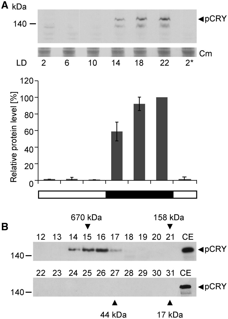
Figure 2.
Analysis of pCRY accumulation in Chlamydomonas over a diurnal cycle and complex formation of pCRY. A, Accumulation of pCRY in Chlamydomonas wild-type cells grown under an LD12:12 cycle. Cells were harvested at the indicated time points. The asterisk indicates the beginning of the next light period at LD2. Equal amounts of proteins from crude extracts (150 μg of protein per time point) were separated by 7% SDS-PAGE and immunoblotted using anti-pCRY antibodies. As a loading control, the PVDF membrane was stained with Coomassie Brilliant Blue R 250 (Cm) after immunochemical detection. From this stain, selected, unspecified protein bands are shown (middle). Quantified pCRY protein levels of three biological replicates are shown at bottom. B, Oligomeric state of pCRY in Chlamydomonas. Wild-type cells were harvested at night (LD22). Crude extracts of soluble proteins (1 mg) were loaded onto a Superdex 200 Increase 10/300 GL size-exclusion column, and 0.5-mL fractions of the elution were collected. Proteins from 100 µL of each fraction (numbers 12–31) were separated by 7% SDS-PAGE along with 100 µg of crude extract (CE) and used for immunoblotting with anti-pCRY antibodies. The molecular masses of the standard protein markers are indicated by arrowheads.

