
Figure 3.
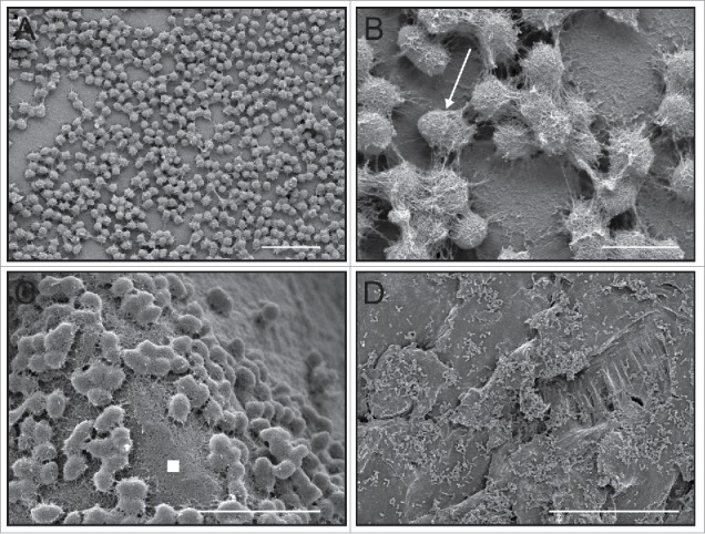
Four previously-unpublished low-voltage scanning electron microscopy (LV-SEM) of E. faecalis microcolony and biofilm formation under a range of on in vitro conditions similar to our previously published work demonstrating both the rapidity in which attachment and extracellular matrix production begins as well as the general structural morphology of the ECM when the matrix is properly preserved using a cationic dye stabilization system. (A) Aclar fluoropolymer coupons,20 24 hrs, CDC biofilm reactor system, bar = 6 µm;51 (B) higher magnification from (A) showing the E. faecalis diplococci surrounded by the sweater-like ECM (arrow), bar = 1 µm; (C) note that the biofilm ECM not only covers cells, but extends into interstitial space between cell clusters (white filled square), regenerated cellulose membrane, 24 hrs, bar = 5 µm;50 (D) polycarbonate coupon, 24 hrs, bar = 50 µm.