Figure 5.

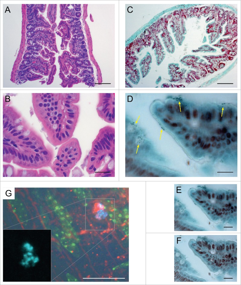
Histological and IFM examination of murine GI tissue from representative sections adjacent to those prepared for SEM.A-F: Histology of the colonized murine terminal ileum. (A). H&E demonstrating normal ileal morphology; note marked lack of obvious inflammatory response. (B). Higher magnification of A. (C). Tissue Gram stain (Hucker-Twort) at low magnification. (D). Higher magnification of C showing numerous small, Gram-positive organisms (yellow arrows showing clusters of small, dark blue cells at the luminal border of epithelial cells. (E). Additional image of same section ∼1 μm deeper in the tissue. (F). Additional image of same section ∼2 μm deeper where the murine epithelium is in focus. G: IFM of the colonized proximal murine colon. (G). Immunofluorescent microscopy of the murine proximal colon colonized by the constitutive CFP-expressing strain OG1RF cfp+. Blue (cyan) = CFP-positive E. faecalis cells; red = Alexa Fluor 594. WGA ; green = autofluorescence from the murine epithelium. The red WGA lectin antibody primarily labels the host epithelial layer as well, but some labeling can be seen around bacterial cells that have begun to produce polysaccharide-rich extracellular matrix. Bars: A, C = 100 µm; B, D - F = 20 µm.
