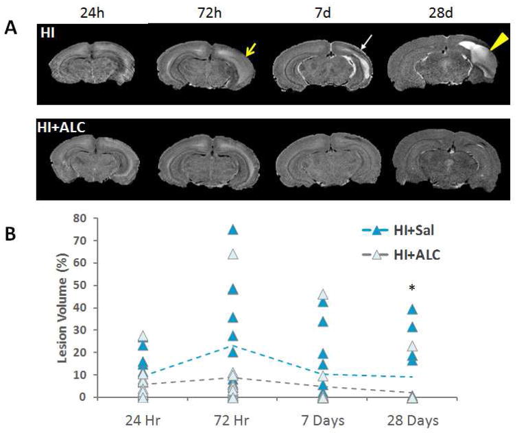
Figure 1.
A) Representative skull-stripped T2-weighted images showing the lesion over time. T2-weighted image for typically developing brain injury and a rat pup 24 hours, 72 hours, 7 days and 28 days after hypoxia ischemia (top panel) and a rat pup treated with ALCAR after hypoxic ischemia (bottom panel). Yellow arrow: edema; white arrow: scar tissue; yellow arrow head: cyst. B) Scatter plot of lesion volume compared to the ipsilateral hemisphere volume. Treatment with ALCAR reduced the size of the lesion in most animals at the final timepoint (28 days post-HI). * denotes significant Kruskall Wallace, p < 0.038.

