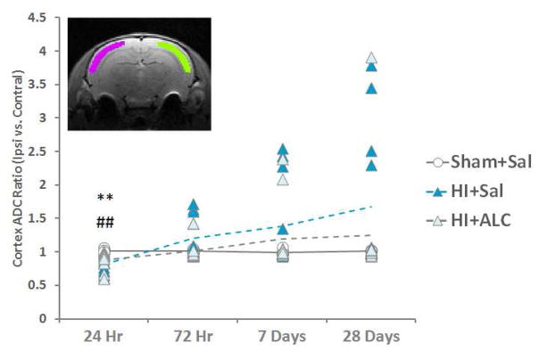
Figure 3.
The cortex ADC ratio (ipsilateral cortex ADC over contralateral cortex ADC) was tested with Kruskal-Wallis test within each time point. Post-hoc analysis was performed with Dunn-Bonferroni test. Decreased diffusivity was observed in HI animals. ALCAR did not prevent this alteration. **=p<0.01 HI vs. Sham; ##=p<0.01 HI+ALCAR vs. Sham. Inset: illustrates the ROIs for ADC measurement (green: ipsilateral cortex; pink: contralateral cortex)

