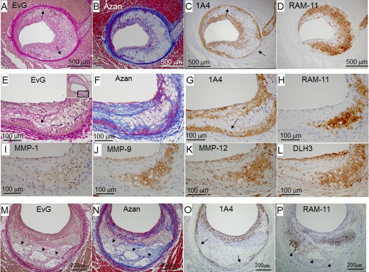
Fig. 4.
Representative photomicrographs of fibroatheromas from early to advanced stages: an early fibroatheroma (female, 19 months old, panels A–D), intermediate lesion between early and advanced fibroatheromas (female, 16 months old, panels E–L), and advanced fibroatheroma (male, 10 months old, panels M–P). Panels A–D, E–L, and M–P are serial sections. Panels F–L are higher magnified views of the square area in panel E. Arrows indicate where the elastic lamina disappeared. Arrowheads indicate necrotic cores. EvG, Elastic van Gieson staining.

