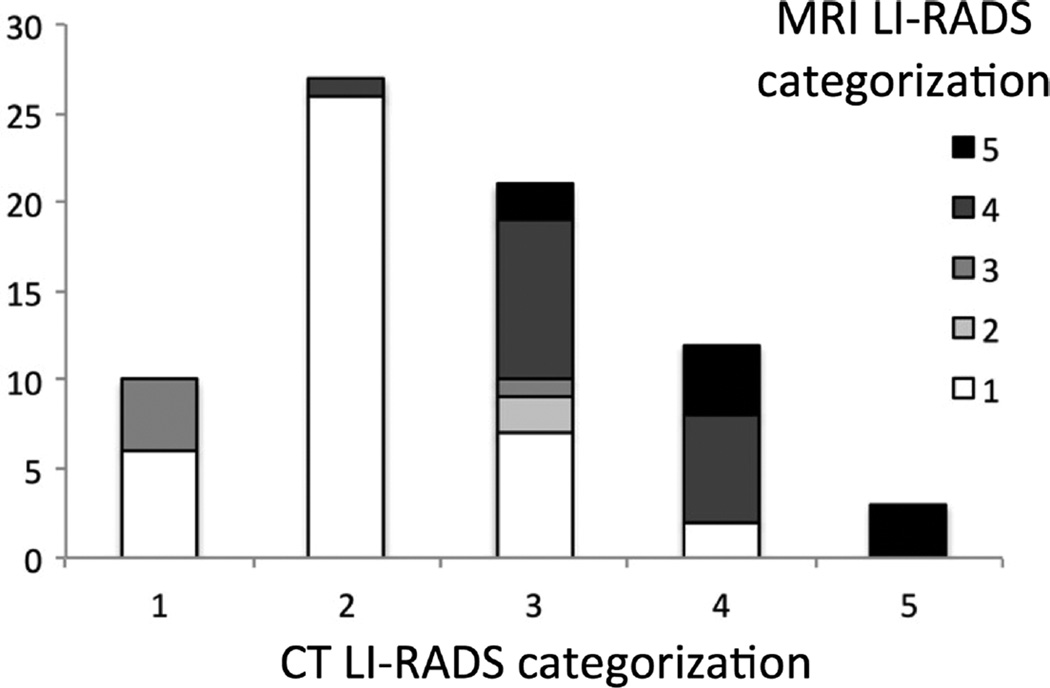
FIGURE 1.
Liver Imaging Reporting and Data System categorization based on CT and MRI characteristics. The most common change in categorization was from LI-RADS 2 on CT to LI-RADS 1 on MRI. Five lesions were originally classified as definitely benign (LR-1) or probably benign (LR-2) and were reclassified as intermediate (LR-3) or likely malignant (LR-4).

