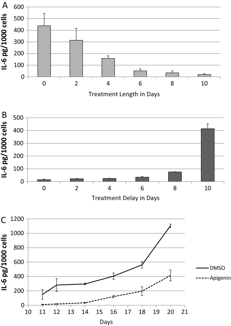
Fig. 3.
Effect of timing of apigenin treatment on IL-6 secretion. a, b HCA2 fibroblasts were seeded at 10,000/cm2 into two 24-well plates and induced to senesce by IR. 10 days later, we varied the length of continuous treatment with apigenin starting immediately after IR in one plate (a), and in the other plate, we varied the day of initiation of treatment with apigenin after IR (b). Duplicate samples were treated with media containing apigenin or DMSO and refreshed every 48 h. On day 9, media were replaced with serum-free media containing apigenin or DMSO, and 24 h later, cells were counted and conditioned media analyzed for IL-6 secretion. c HCA2 fibroblasts were seeded at 10,000/cm2 into a 24-well plate and induced to senesce by IR. Immediately following IR, media were refreshed with DMSO or apigenin and incubated for 10 days (media refreshed every 48 h with DMSO or apigenin). On day 10, cells were washed and incubated with serum-containing media except for the first sample (day 11) that was replaced with serum-free media. Samples for subsequent time points were similarly washed and media replaced with serum-free media 24 h before collection. After the final time point on day 20, CM for all time points were analyzed for IL-6

