FIG 2.
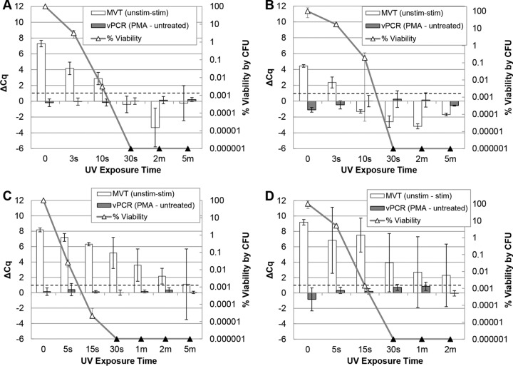
MVT and vPCR of UV-treated A. hydrophila and E. coli. Triplicate cell suspensions in ATW (A. hydrophila) (A and B) or PBS (E. coli) (C and D) at 1 × 10⁸ CFU/ml were separately exposed to a time course of UV irradiation. Equal portions of each time point sample were subjected to MVT (white bars, left axes), vPCR (gray bars, left axes), and plating (triangles, right axes). Culture results (triangles) are displayed as percent viability relative to unexposed cells (time zero). Black triangles indicate no detected colonies (below the limit of detection). MVT (RT-qPCR) and vPCR (qPCR) results are both expressed as ΔCq values (left axes). For MVT, positive ΔCq (unstimulated minus stimulated) values of >1 (dashed lines) indicate viable cells. For vPCR, elevated ΔCq (with PMA minus without PMA) values indicate inactivated cells with compromised membranes.

