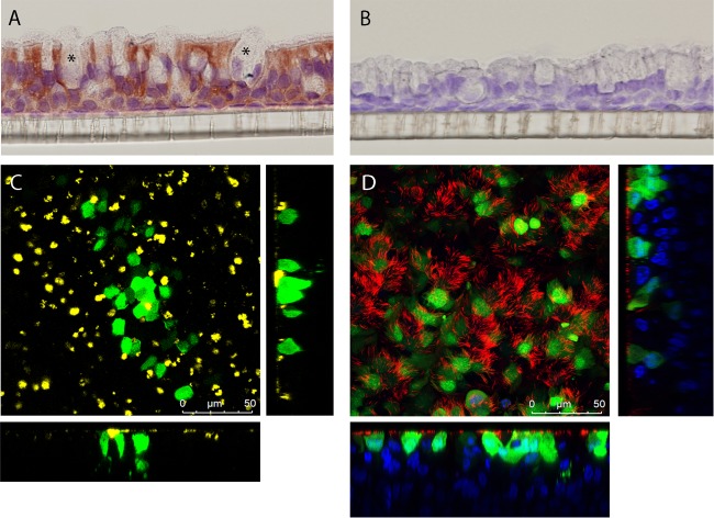
FIG 4.
RSV-A2 infection of primary human airway epithelial cell cultures. (A) Immunohistochemistry staining of CX3CR1 (5 μg/ml, indicated by red staining) on the HAE cell membrane. Asterisks, goblet cells. (B) IgG isotype control staining (5 μg/ml). (C) Top and side views of RSV-A2-infected HAE cells, showing RSV-infected cells in green (GFP), goblet cells in yellow (MUC5B), and nuclei in blue (Hoechst 33342). (D) Top and side views of RSV-A2-infected HAE cells, showing RSV-infected cells in green (GFP), cilia in red (β-tubulin), and nuclei in blue (Hoechst 33342).

