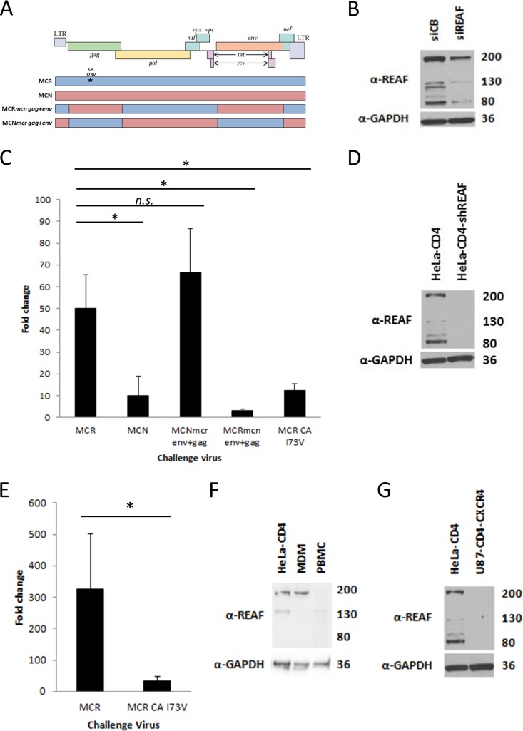
FIG 1.
REAF is an important component of Lv2. (A) A schematic representation of the HIV-2 molecular clones, chimeric viruses, and SDM (*) used to map the determinants of REAF restriction. LTR, long terminal repeat. (B) WB analysis of HeLa-CD4 cell lysate following REAF siRNA knockdown compared with the nontargeting control (siCB). GAPDH was added as a loading control. (C) Titration of constructs on HeLa-CD4 cells transiently transfected with siREAF showing fold changes compared to cells transfected with the siCB control (compared with HIV-2MCR: HIV-2MCN, P = 0.004; HIV-2MCNmcr env+gag, no significant difference; HIV-2MCRmcn env+gag, P < 0.001; HIV-2MCR CA I73V, P = 0.003). (D) WB of REAF knockdown in HeLa-CD4-shREAF cells compared to HeLa-CD4 cells. GAPDH was added as a loading control. (E) Fold changes in HeLa-CD4-shREAF cells infected with HIV-2MCR and HIV-2MCR CA I73V confirm the Lv2 phenotype in the stable knockdown cells (P = 0.016). (F) WB of REAF levels in MDMs and PBMCs compared to those in HeLa-CD4 cells. GAPDH was added as a loading control. (G) WB of REAF levels in U87-CD4-CXCR4 cells compared to those in HeLa-CD4 cells. GAPDH was added as a loading control. Gel run with that shown in panel D. The values to the right of panels B, D, F, and G are approximate molecular sizes in kilodaltons.

