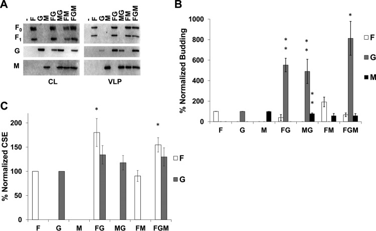
FIG 1.
NiV F and M support incorporation of NiV G into VLPs. (A) Cell lysates (CL) and viruslike particles (VLPs) were isolated from HEK293T cells transfected for 24 h with the corresponding combinations of NiV F, G, and M. Transfections were done with F:G:M ratios of 4:1:1, with pcDNA3.1 as an empty vector. All samples were run through SDS-PAGE and Western blotting. For NiV F, the uncleaved form is designated F0, and part of the cleaved form is designated F1. (B) Densitometric analysis was used to quantify all bands and budding indices were calculated by dividing the band intensity of each VLP band by their corresponding band in CL. This was normalized to the ratio of individually expressed F, G, or M and is shown as a percentage of that ratio. (C) Flow cytometric analysis was used to assess cellular surface expression of F and G and these values were normalized to corresponding single expression values after removal of background. Error bars designate values for standard errors of the mean. Three or more independent experiments were used for each ratio, and the statistical significance was evaluated with one-sample t tests. Statistical significance is indicated by asterisks: *, P < 0.05; and **, P < 0.01.

