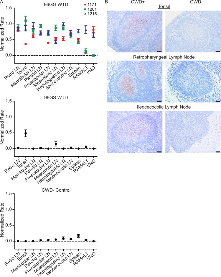
FIG 5.
Increased PrPCWD tissue burden is present in systemic lymphoid tissues in 96GG deer at 4 months postexposure. (A) RT-QuIC PrPCWD amyloid formation was detected in all tissues from at least two of the three 96GG WTD collected at 4 MPE (P < 0.05, unpaired t test). The two tissues that did not have statistically significant rates of amyloid formation were 1201 vomeronasal organ and 1201 RAMALT. Compared with 3-MPE tissues (Fig. 4), 4-MPE tissues displayed higher rates of amyloid formation, consistent with a larger PrPCWD tissue burden. Tonsil from 96GS WTD had detectable amyloid formation at this collection time (P < 0.01, unpaired t test). Data from three 96GG deer (1171, 1201, and 1215), one 96GS deer, and one negative-control deer are represented as means and SEM of 12 replicates from 3 separate experiments. (B) Representative TSA-IHC from 96GG WTD collected at 4 MPE. All lymphoid tissues displayed PrPCWD immunoreactivity in the germinal centers of follicles. The greatest staining was observed in oropharyngeal lymphoid tissues. No staining was observed in corresponding negative-control tissues. IHC images are at ×200 magnification; scale bar, 50 μm.

