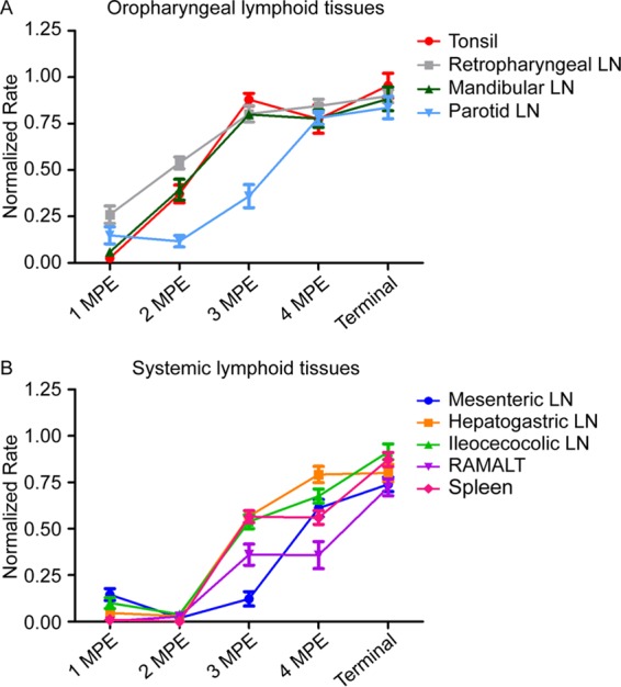
FIG 8.

PrPCWD tissue burden in early infection approximates terminal disease tissue levels in oropharyngeal lymphoid tissues. The prion concentration in oropharyngeal tissues was estimated by averaging the amyloid formation rate from all deer at each collection time point. (A) Estimation of prion replication kinetics in oropharyngeal lymphoid tissues during early CWD infection. In oropharyngeal lymph nodes, the tonsil and retropharyngeal and mandibular lymph nodes displayed a rapid PrPCWD accumulation and reached tissue PrPCWD levels comparable to that for terminal disease burden by 3 MPE (3 MPE versus terminal, P > 0.05, unpaired t test). The parotid lymph node displayed a slightly slower prion accumulation but reached prion levels comparable to that of terminal disease by 4 MPE (4 MPE versus terminal, P > 0.05, unpaired t test). (B) Estimation of prion replication kinetics in gastrointestine-associated lymphoid tissues (GALT) and systemic lymphoid tissues during early CWD infection. PrPCWD seeding activity was not detected in distal GALT and the spleen until 3 MPE. At 4 MPE, GALT and spleen had statistically significantly lower rates of amyloid formation than corresponding terminal tissues (4 MPE versus terminal, P < 0.01, unpaired t test), indicating these lymphoid tissues did not reach maximum prion burdens during the time course of our study.
