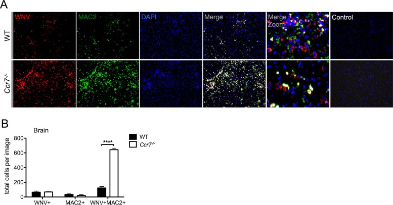
FIG 6.
Ccr7 restricts the number of WNV-infected myeloid cells in the CNS. (A) Paraffin-embedded brain sections from WT and Ccr7−/− mice on day 12 postinfection were stained for WNV (red), MAC2 (green), and nuclear 4′,6′-diamidino-2-phenylindole (DAPI; blue). WNV-infected myeloid cells costained positive for both WNV and MAC2 and appear in yellow. Control conditions, under which only secondary antibodies were used, are shown. Representative images are shown at a magnification of ×10, with zoomed-in panels. (B) Quantification of single- and double-positive WNV and MAC2 cells from n = 3 images per mouse strain in matching brain areas. Data are shown as means ± standard deviations. ****, P < 0.0001.

