FIG 2.
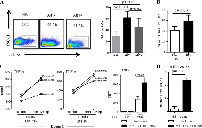
miR-126-5p upregulates TNF-α production in LPS-stimulated primary monocytes. (A) HIV-1 infection resulted in increased TNF-α production of monocytes in response to LPS stimulation. Examples of TNF-α production of monocytes with and without LPS stimulation are shown on the left. PBMCs from the HIV− (n = 9), ART− (n = 8), and ART+ (n = 8) groups were stimulated with LPS (100 ng/ml) for 20 h. TNF-α production in monocytes was quantified by gating CD14−-positive cells in a flow cytometry-based intracellular staining assay and statistically calculated by groups. TNF-α production is significantly higher in the ART− (P < 0.0001) and ART+ (P = 0.02) than in the HIV− group. ART treatment significantly reduces TNF-α production (P = 0.03 for the ART− group versus the ART+ group). (B) TNF-α production is significantly upregulated in nonclassical monocytes. TNF-α production was quantified as described above; nonclassical monocytes were gated as CD14+ CD16++, and TNF-α production in this subset was compared between the HIV-1-seronegative and HIV-1-infected groups. (C) Overexpression of miR-126-5p in primary monocytes derived from the HIV− subjects profoundly enhances TNF-α production. Here, 1 × 10⁵ transfected monocytes were stimulated with LPS (100 ng/ml) for 20 h, and the supernatants were collected for TNF-α analysis. The level of TNF-α was quantified by ELISA. The results shown on the left were from three independent experiments in one donor, and the production of TNF-α in response to LPS stimulation in three donors is shown on the right. (D) Effective transfection of miR-126-5p mimic in primary monocytes was observed. Primary monocytes derived from the HIV− group were electrotransfected with control or miR-126-5p mimic, and the transfection efficacy was determined by qPCR.

