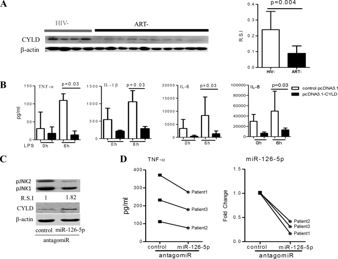
FIG 4.
miR-126-5p/CYLD/JNK axis regulates the responses of monocytes to LPS stimulation during chronic HIV-1 infection. (A) HIV-1 infection resulted in a reduction in CYLD protein. Western blot analyses of CYLD protein in monocytes from ART− individuals (n = 10) were compared with that from HIV− individuals (n = 8), shown as an example (left) and as R.S.I. (right). (B) Overexpression of CYLD in primary monocytes (n = 5) derived from the ART− group suppresses their inflammatory responses to LPS stimulation. The supernatants were collected at 6 h after LPS (100 ng/ml) stimulation of 5 × 10⁴ primary monocytes transfected with pcDNA3.1-CYLD (codon optimization) plasmids or pcDNA3.1 plasmids for 72 h. The levels of TNF-α, IL-6, IL-1β, and IL-8 were quantified using BioLegend LEGENDplex CBA. (C) Inhibition of miR-126-5p increases CYLD protein expression and decreases JNK phosphorylation in monocytes from the ART− group. Expression of CYLD protein and JNK phosphorylation were analyzed by Western blotting in primary monocytes that were electrotransfected with control antagomir or miR-126-5p antagomir for 72 h. Transfection of the miR-126-5p antagomir resulted in an increase in CYLD protein levels and a decrease in phosphorylated JNK levels. (D) Downregulation of miR-126-5p in monocytes from the ART− group resulted in decreased TNF-α production. Monocytes from the ART− group (n = 3) were electrotransfected with either the control antagomir or the miR-126-5p antagomir; supernatants were collected at 12 h after LPS (100 ng/ml) stimulation of 5 × 10⁴ transfected monocytes. The downregulation of miR-126-5p was detected by qPCR and is shown on the right.

