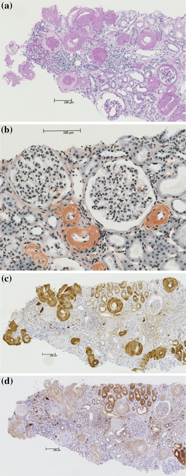
Fig. 1.

Light microscopy findings. a Periodic acid Schiff (PAS) staining, showing thickening of the walls of the small arteries and arterioles with weakly PAS-positive material, and interstitial invasion by lymphocytes. b Direct fast scarlet staining, showing positive staining of the vascular walls and partial positive staining of the glomeruli including sclerotic glomeruli. c Immunohistochemical staining for kappa chains, showing positive staining of the vascular walls and partial positive staining of the glomeruli including sclerotic glomeruli. d Immunohistochemical staining for lambda chains
