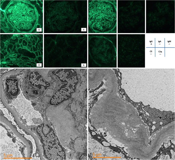
Fig. 2.
Upper panel shows immunoreactivity for κ, λ, IgG, IgA, IgM, C3, and C1q (magnification, ×400). Linear and circumferential faint IgG and κ immunoreactivity were observed along with the glomerular capillaries and tubular basement membranes. The Lower panel shows an electron microscopic image (magnification, ×50,000). No electron-dense deposition is present at the glomerular subendothelial cells. Glomerular basement membrane thickening was predominant and had an average width of 847 nm

