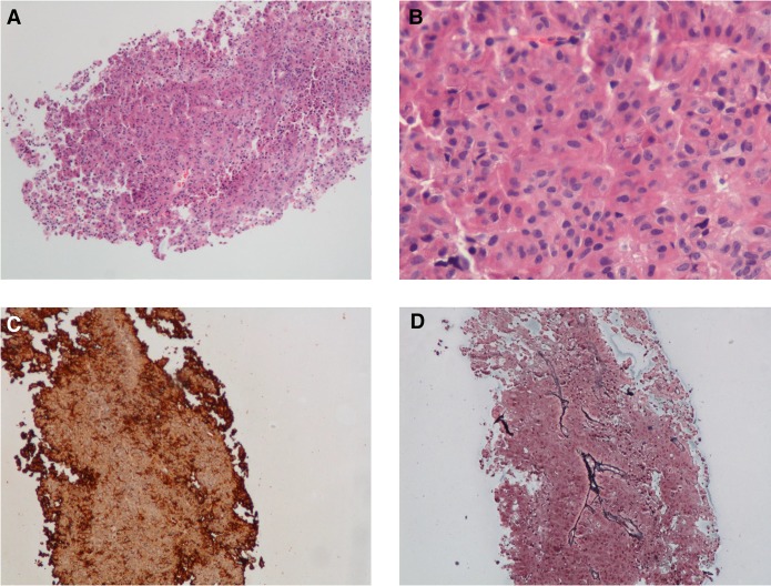
Figure 2.
Histologic sections revealed a monotonous tumor composed of medium-sized cells with finely stippled chromatin (A,B). Tumor cells were strongly positive for adenocorticotropic hormone (ACTH) by immunohistochemistry (C), whereas a reticulin stain (D) showed effacement of the fibrovascular septae. (A) Hematoxylin and eosin (H&E) 100×; (B) H&E 400×; (C) ACTH immunostain, 100×; (D) reticulin, 100×.

