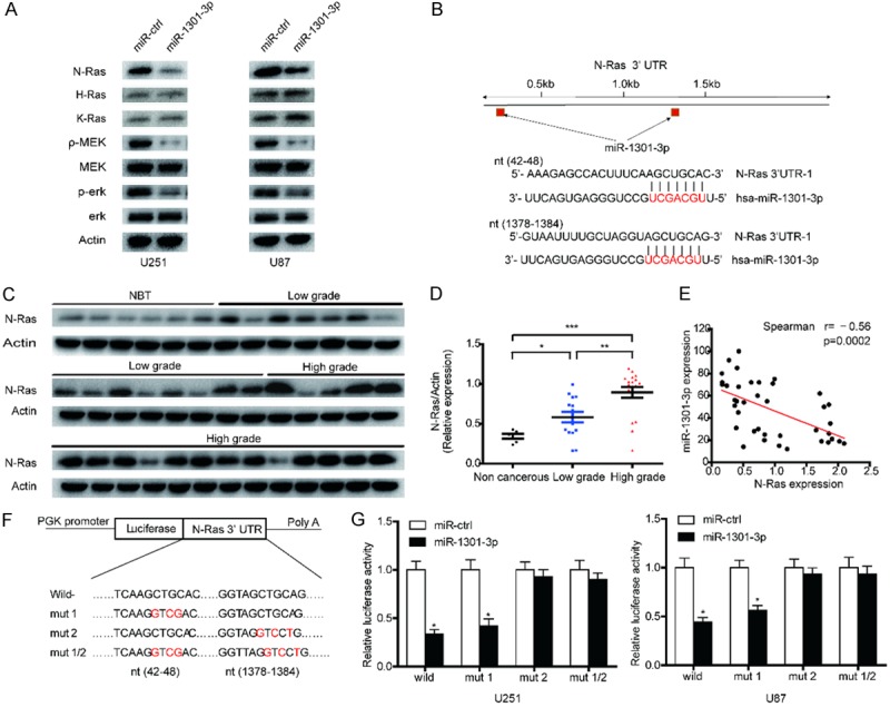
Figure 3.

MiR-1301-3p directly targets N-Ras and suppresses the activity of the Ras-MEK-ERK1/2 pathway in GBM cells. A. N-Ras, H-Ras, K-Ras, MEK, p-MEK, ERK1/2 and p-ERK1/2 expression levels in indicated cells were determined by western blotting. B. Predicted miR-1301-3p binding sites in the 3’-UTR of the N-Ras gene. C, D. The expression levels of N-Ras in NBTS and glioma specimens were determined by western blotting; the fold changes were normalized to β-Actin. The non-neoplastic brain tissues (n=6) were collected from brain trauma surgery. The low-grade (n=15) represents samples derived from grades I and II glioma tissues, whereas high-grade (n=18) represents grades III and IV glioma tissues. Data represent the means ± SD from three independent experiments. *P<0.05, **P<0.01, ***P<0.001. E. Pearson’s correlation analysis of the relative expression levels of miR-1301-3p and the relative protein levels of N-Ras. F. Wild-type and mutant N-Ras 3’-UTR reporter constructs. G. Luciferase reporter assays were performed in U251 and U87 cells with co-transfection of indicated wild-type or mutant 3’-UTR constructs and miR-1301-3p mimic. The data shown are representative of three independent experiments. Data shown are mean ± SD of three independent experiments. *P<0.05.
