Figure 2.

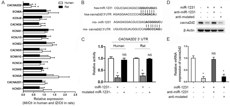
miR-1231 suppresses cacna2d2 expression by targeting its mRNA. (A) The mRNA level of cacna2d2 was significantly decreased in both human and rat ischemic heart samples. Real-time PCR analysis of the expression of predicted 14 ion channel target genes in ischemic hearts compared with the healthy control heart samples in both human and rats as depicted in Figure 1. In human samples, the randomly selected three pairs of human control non-ischemic hearts and MI hearts were compared, and in rat samples, three randomly chosen pairs of NIZ, BZ, IZ and Ctl samples from rats at 14 days post-MI were compared as well. Results of MI or IZ relative to Ctl were shown (*, P<0.05 compared with Ctl group, n=3). (B) Schematic illustration of sequence complimentarity between miR-1231 and the 3’-UTRs of cacna2d2 mRNAs in human (hsa) and rat (rno), provided with computational and bioinformatics-based approach using TargetScan [2]. Watson-Crick complementarity was presented in bold text and linked with its paired nucleotide. (C) 3’-UTR of human or rat cacna2d2 was target for human or rat miR-1231 in HEK293 cells, respectively. HEK293 Cells were transiently transfected with luciferase reporters linked to the 3’-UTR sequences of human or rat CACNA2D2 gene as indicated. The luciferase reporter activity was measured after 1 day co-expression. Human or rat miR-1231 repressed luciferase reporter gene activity, however, mutatedmiR-1231 was unable to decrease luciferase activity. Mean ± SD; n=8; *P<0.05; NS, not significant compared with Ctl). Unpaired Student’s t-test. (D, E) The mRNA and protein expression of cacna2d2 were downregulated by miR-1231 in cultured myocytes. The protein (D) and mRNA (E) levels of cacna2d2 were determined by Western blotting and qRT-PCR in cultured neonatal rat cardiac myocytes, which were transfected with miR-1231 alone or in combination with anti-miR-1231or anti-mutated. Left, β-Actin was used as a loading control and representative results of Western blotting bands were shown; Right, mRNA levels in four groups relative to Ctl were shown (*, P<0.05; NS, not significant compared with Ctl group, n=3).
