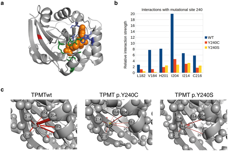
Figure 6.
(a) Three-dimensional structure of human TPMT with the Y240 position in the β9-sheet marked in red. The bound coproduct of SAM, S-adenosylhomocysteine (SAH) is showed as spheres (orange) together with the SAH interacting residues (blue). Residues involved in binding the substrate 6-MP are showed in green. (b) Analysis of the relative interaction strength for positions interacting with position 240 from the MD simulations of WT, Y240C, and Y240S. (c) Relative interaction strengths visualized in the three-dimensional structure as red rods with a thickness proportional to the interaction strength for TPMTwt, TPMT p.Y240C and TPMT p.Y240S respectively. For clarity only the interactions with a relative strength larger than 1.0 are included.

