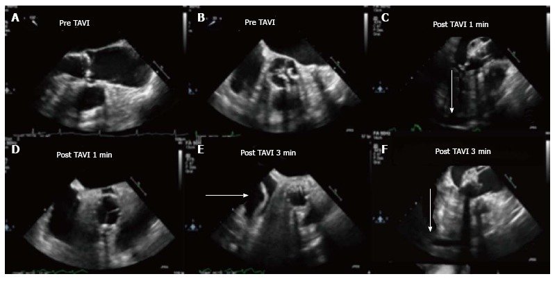
Figure 1.

Aortic root rupture and aortic root hematoma (Case 1). A and B: Aortic valve assessment before TAVI, TEE mid esophageal long axis view (A) and short axis view (B); C and D: Aortic valve assessment post TAVI 1 min; E and F: Aortic valve assessment post-TAVI 3 min. We can observe the development of the aortic root hematoma and the pericardial effusion (arrowhead). TAVI: Transcatheter aortic valve implantation; TEE: Trans-esophageal echocardiography.
