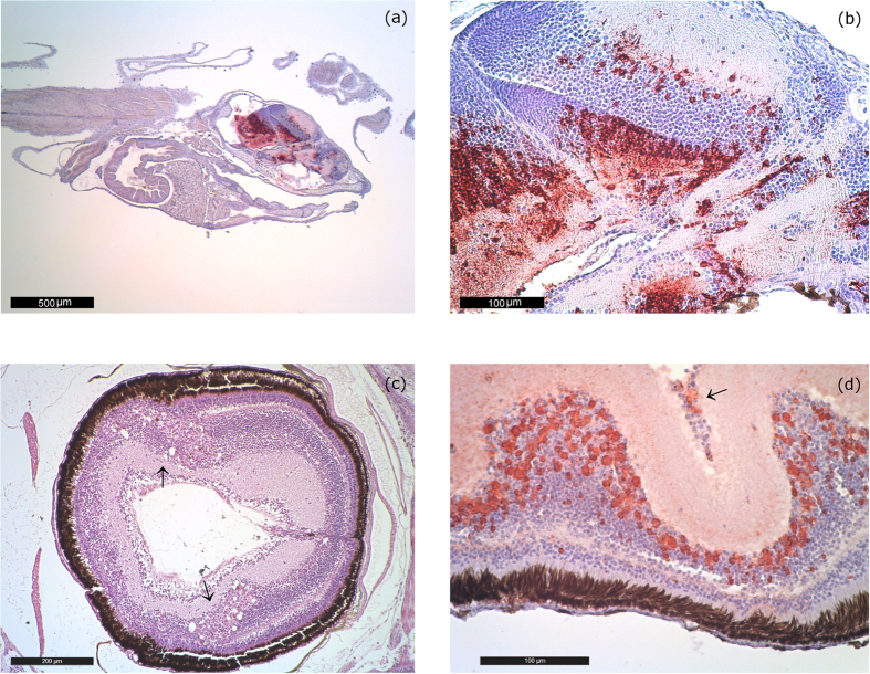
Figure 1. Acute lesions.
(a) IHC Sea bream larvae of 16 days of age from farm 1 (2014–15). Bright red immunoprecipitates are visible in the telencephalon, mesencephalon, diencephalon (hypothalamus) and cerebellum. IHC labeling is generally higher in larvae and post larvae than in juveniles. 40 magnification. (b) IHC of 16 day-old larvae from farm 1 (2014–15). Massive immunoprecipitates in the telencephalon, mesencephalon, diencephalon (hypothalamus). Remarkably, no vacuolization is noticeable. 250 magnification. (c) Sea bream postlarvae of 45 days of age from farm 1 (2014–15). Vacuolation in the inner nuclear layer of the retina (arrow). H&E 100 magnification. (d) IHC of a 55 day-old seabream eye collected in farm 1 (2014–15). Immunoprecipitates are evident in the inner nuclear layer and the ganglion cell layer (arrow) of the retina. 250 magnification.

