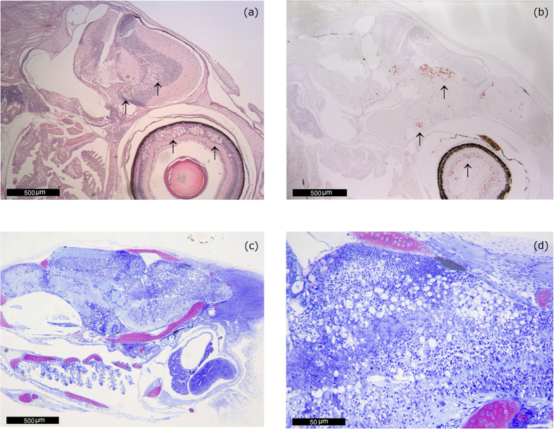
Figure 3. Lesions in past VER outbreaks in sea bream.
(a) VNN-associated damage in the encephalon and in the retina (arrows) of a 45 day-old seabream from samples collected during the Cyprus outbreak in 2010. H&E. 40 magnification. (b) IHC of a 45 day-old seabream brain from another specimen collected during the Cyprus outbreak (2010). Immunoprecipitates are visible in the telencephalon (olfactory lobes, preoptic nucleus) in mesencephalon (tectum opticum and tegmentum) and retina (arrows). 40 magnification. (c) 43 day-old Sea bream postlarvae collected during the 2005 outbreak in Portugal. Acute, severe and extensive lesions. Extensive vacuolation is noticeable in the telencephalon, mesencephalon, diencephalon (hypothalamus) and medulla oblongata. Methacrylate section. Toluidine Blue stain (TB). 40 magnification. (d) 43 day-old Sea bream postlarvae collected during the 2005 outbreak in Portugal. Higher magnification of the lesions observed in (c). Notice the presence of a large number of cells with large vacuoles within the cytoplasm in the metencephalon and also in the mesencephalon. Methacrylate section. Toluidine Blue stain (TB). 400 magnification.

