
Fig. (2).
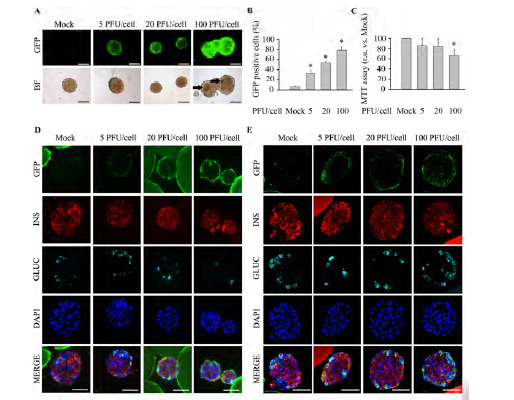
High pHRSIN DUAL-GFP PFU/cell levels compromise islet viability with sub-optimal islet transduction efficiency. Freshly isolated murine islets were exposed to increasing PFU/cell of pHRSIN DUAL-GFP. Non-transduced islets (Mock) were used as control. (A) Representative images of ex vivo cultured entire live transduced islets. Top; GFP expression was assessed by fluorescence acquisition using an ImageXpress Microsystem. Low; Bright field images. Images were captured at 4 days post-infection. Arrows indicate necrotic areas. Scale-bars indicate 100 µm. n=4 experiments per condition. (B) Transduction efficiency, defined as the percentage of islet cells expressing GFP, was determined by flow cytometry in disaggregated islets at 4 days post-infection. n=6 per condition. (C) Determination of islet metabolic activity using the MTT assay at 4 days post-infection n=4-6 per condition. (D-E) Representative immunofluorescence images of Affi-Gel bead-embedded pancreatic islets 4 days post-infection. Antibodies against GFP (green), insulin (red) and glucagon (cyan) were employed. Of note, in some instances the Affi-Gel beads, emitted a non specific fluorescent signal along with GFP (Green) and insulin (red). (D) Wide-field fluorescence microscopy. (E) Confocal microscopy. Scale-bars, 50 µm. n=3 per condition. Data are represented as the mean ± SEM. * p< 0.05 versus control non-transduced islets.