
Fig. (4).
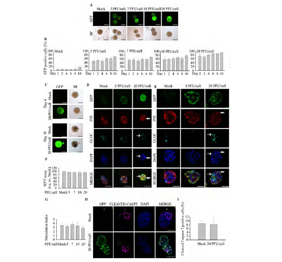
Mild trypsinization combined with 20 PFU/cell represents the optimal infection protocol for murine islets. Freshly isolated murine islets were treated with 0.5 X trypsin-EDTA (250 mg/l; 0.48 mM EDTA) and subsequently exposed to increasing PFU/cell of pHRSIN DUAL-GFP. (A) Representative images of GFP fluorescence emitted from live islets: Top; GFP expression was assessed by fluorescence acquisition using an ImageXpress Microsystem. Low; Bright field images. Images were captured at 4 days post-infection. Scale-bars 100 µm. n=4 experiments per condition. (B) Transduction efficiency in 0.5 X trypsin-EDTA treated islets at different days after transduction was determined by flow cytometry in dispersed islets. n=4 per condition. (C) Representative images of live islets exhibiting GFP fluorescence 4 and 10 days post-treatment: Top; GFP expression was assessed by fluorescence acquisition using an ImageXpress Microsystem. Low; Bright field images. Scale-bars 100 µm. n=4 experiments per condition. (D-E) Representative immunofluorescence images of Affi-Gel bead-embedded pancreatic islets trypsin-treated and transduced or not with pHRSIN DUAL-GFP. Antibodies against GFP (green), insulin (red) and glucagon (cyan) were employed. Images were captured in samples fixed at 4 days post-infection using either wide-field fluorescence microscopy (D) or confocal microscopy (E). Of note, in some instances the Affi-Gel beads, emitted a non specific fluorescent signal along with GFP (green) and insulin (red). Filled arrows indicate transduced cells expressing insulin; Non-filled arrows indicate transduced cells expressing glucagon. Scale-bars 50 µm. n=3 per condition. (F) Determination of islet metabolic activity subsequent to a 0.5 X trypsin-EDTA treatment followed by transduction with 20 PFU/cell. A MTT assay was performed 4 days post-infection. n=3-4 per condition. (G) Glucose-stimulated insulin secretion was assessed in islet treated with 0.5 X trypsin-EDTA followed by transduction with increasing amount of pHRSIN DUAL-GFP. (H) Representative immunofluorescence images of Affi-Gel bead-embedded pancreatic islets 0.5X trypsin-treated and transduced or not with pHRSIN DUAL-GFP. Antibodies against GFP (green) and cleaved Caspase 3 (magenta). Images were captured in samples fixed at 4 days post-infection using confocal fluorescence microscopy. n=3 per condition. (I) Determination of apoptosis rate by quantification of cleaved caspase 3 positive cells in islets 0.5X trypsin-EDTA-treated and transduced or not with pHRSIN DUAL-GFP. n=3 per condition. Data are represented as the mean ± SEM of n=3. * p < 0.05 versus control non-transduced 0.5 X trypsin-EDTA treated islets.