
Fig. (5).
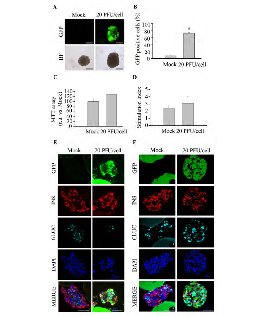
Human islets are efficiently transduced using the optimized protocol. Human islets obtained from cadaveric donors were initially treated with 0.5 X trypsin-EDTA (250 mg/l trypsin; 0.48 mM EDTA) and then transduced with pHRSIN DUAL-GFP at 20 PFU/cell. (A) Live imaging reveals GFP expression in human islets 4 days post-infection: Top; GFP expression, assessed by fluorescence acquisition using an ImageXpress Microsystem, Bottom; Bright field images. Scale-bars 100 µm. n=3 per condition. (B) Transduction efficiency in 0.5 X trypsin-EDTA treated islets was determined by flow cytometry of dispersed islets at 4 days post-transduction with 20 PFU/cell. n=3 per condition. (C) Islet metabolic activity was assessed using the MTT assay. n=3 per condition. (D) Glucose-stimulated insulin secretion was assessed in either control islets or islet treated with 0.5 X trypsin-EDTA followed by transduction with 20 PFU/cell of pHRSIN DUAL-GFP. n=3 per condition. (E-F) Co-immunostaining of GFP (green), insulin (red) and glucagon (cyan) was performed on sections from Affi-Gel bead-embedded human pancreatic islets subsequent to treatment. Images were captured in samples fixed at 4 days post-infection using wide-field fluorescence microscopy (E) or confocal microscopy (F). Scale-bars 50µm. n=3 per condition. Data are represented as the mean ± SEM. * p < 0.05 versus control non-transduced 0.5 X trypsin-EDTA treated islets.