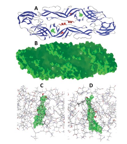
Fig. (4).

Dengue virus envelope protein. (A) Ribbon structure of DENV envelope protein (PDB 1OKE [31]). The cavity pores in each monomer are shown in green. (B) Solid structure of DENV envelope protein (PDB 1OKE). (C) Lowest-energy docked pose of canniflavin A with monomer A of the dimeric envelope protein (PDB 1OKE). The cavity pore is shown in green; hydrogen-bonds are shown as blue dashed lines. (D) Lowest-energy docked pose of paratocarpin L with monomer B of the dimeric envelope protein (PDB 1OKE).
