Figure 1.
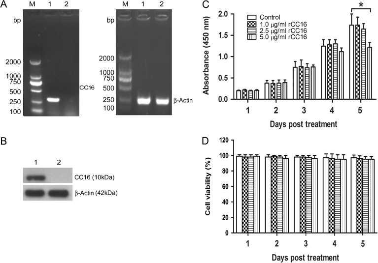
Endogenous expression of CC16 in RAW264.7 cells and effects of rCC16 protein on RAW264.7 cell viability (A,B) The endogenous CC16 expression in RAW264.7 cells was detected by RT-PCR (A) and western blot analysis (B). Lane M: DNA marker; Lane 1: positive control (mouse lung sample); Lane 2: RAW264.7 cells sample. β-Actin was used as an internal control. Each assay was repeated three times with similar results. (C,D) RAW264.7 cells were treated with 1.0, 2.5, or 5.0 μg/ml rCC16 or the same volume of PBS for 1–5 days, and cell proliferation and cell viability were determined by CCK-8 assays (C) and Trypan Blue staining (D), respectively. The data are presented as the mean ± SD of three independent experiments. *P < 0.05 relative to vesicle controls. RT-PCR, reverse transcriptase-polymerase chain reaction, DNA, deoxyribonucleic acid.

