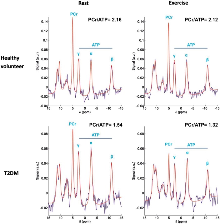
Figure 2.
Rest and exercise myocardial 31P-MR spectra in a healthy volunteer (top row) and a T2DM patient (bottom row). T2DM was associated with significantly lower myocardial PCr/ATP than control at rest, and the decrease was exacerbated during exercise, suggesting a pre-existing myocardial energy deficit in type 2 diabetes mellitus. (Reprinted with permission).

