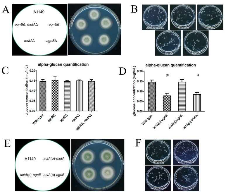
Figure 3.
AgnB and MutA are functional α-glucanases. (A) Freshly harvested conidia (105) of each strain were inoculated on complete medium and the plates were incubated at 30 °C for 48 h. All constructed strains showed the wild-type colony phenotype on solid medium; (B) freshly harvested conidia (5 × 10⁷) were inoculated in a flask with 20 mL complete medium, then the flask was incubated at 30 °C, 150 rpm overnight. All strains behaved the same as the wild-type; (C,D) spores of the indicated strain (2 × 10⁷) were inoculated in 100 mL complete medium. Samples were grown in flasks at 30 °C with 150 rpm for 24 h. α-glucan was extracted from 1 mg of dry cell wall, and then digested to glucose and quantified using an anthrone assay [24]. Results represent the mean of three independent quantification tests with duplicates each time ± standard deviation. The data for each strain were compared with the wild-type (column 1) individually by a Mann Whitney U test. Significant difference (p < 0.05) was indicated by asterisks; (E) conidia of each strain were prepared and inoculated as in (A). Only actA(p)-agnB showed pigment deficiency; and (F) all strains behaved the same as the wild-type in shaken liquid medium. Growth condition was the same as described in (B).

