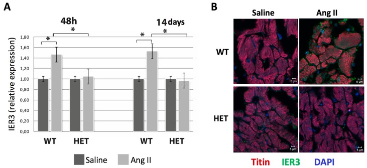
Figure 2.
Impaired cardiac IER3 activation in the titin-deficient mice during heart failure development. (A) Real-time semi-quantitative polymerase chain reaction (PCR) analysis of cardiac IER3 expression in WT and HET Ttn knock-in mice 48 h and two weeks after Ang II infusion (n = 8, * p < 0.05). IER3 response is blunted in the HET mice; (B) Representative IER3 immunofluorescence staining in WT and HET Ttn knock-in mice 48 h after Ang II infusion. Titin staining (red) was used to label the myocardium, nuclei were counterstained with DAPI (blue). Scale bar: 5 μm.

