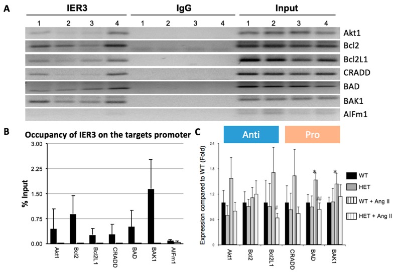
Figure 3.
IER3 targets promotor regions of apoptosis genes in the murine heart. (A) Chromatin immunoprecipitation-PCR (ChIP-PCR) analysis of IER3 target promotor regions. IgG ChIP samples were included as negative control. AIFm1 is a non-related negative control. Heart lysates from four different animals (1–4) were analyzed; (B) Percentages of IER3 occupation on its targets promoter regions. Percentage were calculated as the ratio of occupation of ChIP products to input (p < 0.05 for target promotor sites compared to control); (C) Real-time PCR analysis of IER3 target genes in mouse hearts infused with Ang II for two weeks. The expression of BAD and BAK1 was significantly upregulated in the WT mice with Ang II infusion when compared to the WT with saline infusion, the expression of Bcl2L1 and BAD in the HET mice with Ang II infusion was significantly reduced when compared to the WT mice with Ang II infusion (n = 4 for each group, * p < 0.05 compared to the wildtype mice infused with saline. # p < 0.05, ## p < 0.01 compared to wildtype mice infused with Ang II).

