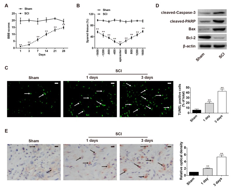
Figure 1.
Establishment and verification of a laboratory rat SCI model. In the SCI group of rats, the skin was incised along the midline of the back, and the vertebral column was exposed. A laminectomy was performed at the T10 level. (A) Basso, Beattie, and Bresnahan (BBB) scores for hindlimb locomotion in each experimental group (n = 4 per group). (B) Quantification of lesion size within the injury site, and measurement of the distance between points 1600 μm rostral and caudal to the epicenter, seven days post-injury. Compared with the sham group, the SCI group had larger spared tissue areas at multiple distances from the lesion epicenter (n = 4/group). (C) TUNEL staining of spared tissues to analyze neuronal apoptosis at one and three days after SCI (n = 3/group/time point). Bright green dots indicate apoptosis positive cells. The white arrows point out TUNEL-positive cells. (D) Protein expression of cleaved caspase-3, cleaved PARP, Bcl-2, and Bax in the spinal cord segment at the contusion epicenter (n = 4/group). β-actin was used as the loading control and for band density normalization. (E) Expression of cleaved caspase-3 after SCI as detected by immunohistochemistry staining in spinal cord tissues collected from each group (n = 3/group/time point). The black arrows indicate cells stained positive with anti-cleaved caspase-3. Data are means ± SEM. ** p < 0.01 vs. the sham group. Scale bars in (C,E) 20 μm.

