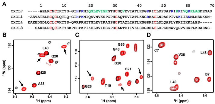
Figure 1.
NMR (nuclear magnetic resonance) characterization of CXCL7 heterodimers. (A) Sequence alignment of platelet-derived CXC chemokines. GAG (glycosaminoglycans)binding residues identified from this study are in blue, dimer interface residues for CXCL7 are in green, and conserved Cys residues are in red; (B–D) Sections of the 1H–15N HSQC (heteronuclear single quantum coherence) spectra showing the overlay of CXCL7 in the free (black) and in the presence of CXCL1 (B, red), CXCL4 (C, red), and CXCL8 (D, red). Arrows indicate new peaks corresponding to the heterodimer. No new peaks were observed in the case of the CXCL8 titration.

