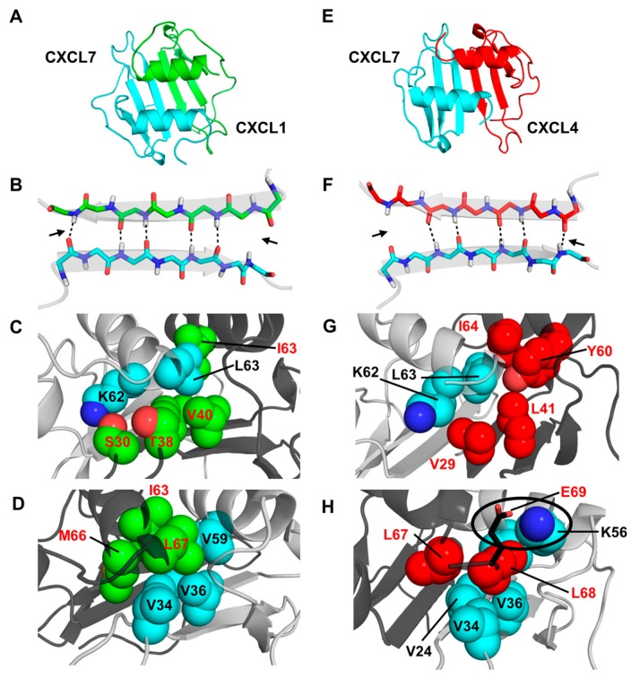
Figure 3.
Structural features of the CXCL7 heterodimers. (A,E) Snap shots of the structural models of CXCL7-CXCL1 and CXCL7-CXCL4 heterodimers from the last 5 ns of the MD (molecular dynamics) simulations; (B,F) A schematic showing the β1-strand dimer interface H-bonds (dashed line) from the final 5 ns of the MD run. Arrows indicate transient H-bonds; (C,D) Packing interactions involving CXCL7 helical (cyan) and CXCL1 β-sheet residues (green) and CXCL1 helical (green) and CXCL7 β-sheet (cyan) residues; (G,H) Packing interactions involving CXCL7 helical (cyan) and CXCL4 β-sheet (red) residues and the CXCL4 helical (red) and CXCL7 β-sheet (cyan) residues. The circle highlights the potential ionic interaction between CXCL4 E69 and CXCL7 K56. Nitrogen atoms are shown in the conventional dark blue and oxygen in light red.

