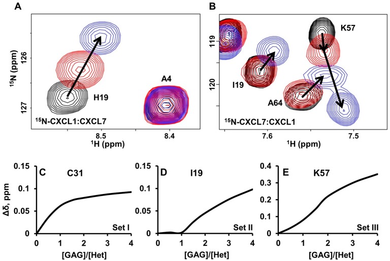
Figure 7.
NMR characteristics of trapped heterodimer-heparin interactions. Sections of the 1H-15N HSQC spectra showing the overlay of CXCL7-CXCL1 trapped heterodimer in the free (black) and heparin dp8 bound form at 1:1 (red) and 1:4 (blue) molar ratios. Arrows indicate the direction of movement. (A) For CXCL1, only linear chemical shifts are observed; (B) In the case of CXCL7, both non-linear chemical shifts (K57) and delayed linear chemical shifts (I19 and A64) are observed; (C–E) Plots of binding-induced chemical shift changes on adding heparin. For CXCL7, (C) hyperbolic, (D) hyperbolic after a delay, and (E) sigmoidal profiles are observed.

