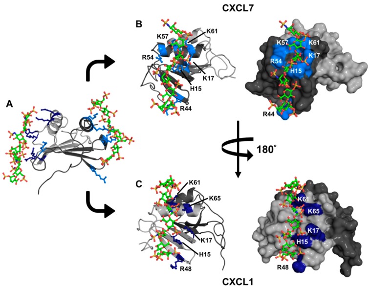
Figure 8.
A model of the heparin-bound CXCL7-CXCL1 heterodimer complex. (A) Ribbon diagram showing that heparin binds to both monomers of the heterodimer. CXCL7 is shown in dark gray and CXCL1 light gray; (B,C) Cartoon and surface plots showing side views of the CXCL7 and CXCL1 monomer faces interacting with heparin dp8, respectively. The basic residues involved in binding are labeled and shown in blue.

