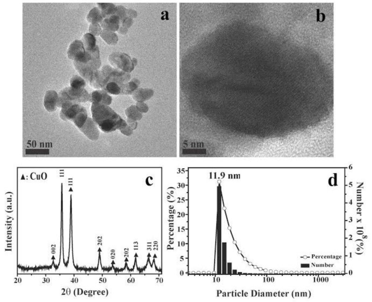
Figure 1.
Shape, crystal structure, and particle size characterization of CuO nanoparticles (NPs). (a,b) scanning electron microscopy (SEM) images of CuO NPs used in this study; black bars: (a) 50 nm and (b) 5 nm; (c) X-ray diffraction (XRD) patterns of synthesized CuO NPs; (d) Size distribution histograms of CuO NPs. The data are representative of two separate experiments.

